Cell Wall Definition
- Around the inner plasma membrane, an outermost layer provides protection and strength to the cell. This outermost layer is the cell wall.
- Protoplast, a living structure, forms the non-living cell wall.
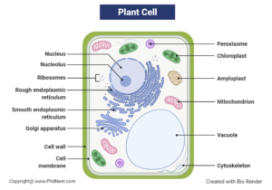
- The cell wall is not a characteristic of all types of cells. Some cells show their presence like plant cells, fungi, algae, and bacteria and are absent in some like animal cells.
- If we look at the composition of the plant cell wall, it is made up of some polysaccharides like cellulose, hemicelluloses, pectin, and protein. While in the fungus cell wall, chitin is the main element. Similarly, bacterial cell walls contain peptidoglycan in them. On the whole, the composition of cell walls varies in different types of cells.
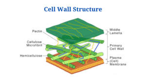
Cell Wall Structure
The cell wall is a rigid structure. This rigidity is due to the covering of a polysaccharide layer that helps in maintaining the cell shape and gives protection.
How to increase Brain Power – Secrets of Brain Unlocked
The Plant Cell Wall
- The unique feature of the plant cell wall is that it is multi-layered and is further divided into three main parts or layers named as the middle lamella, primary cell wall, and the secondary cell wall.
- Middle lamella and the primary cell wall are present in all plant cells, but you will not see secondary cell walls in all plant cells.
- Middle lamella:
This is the outer layer composed of pectin, which is a polysaccharide and thus helps bind cells to one another.
- Primary cell wall:
In between the middle lamella and plasma membrane, the primary cell wall is present. This cell layer is mostly found in young growing plant cells and is composed of polysaccharides like cellulose, hemicelluloses, and pectin. The primary cell wall helps the younger plant cells in their growth by providing flexibility and support.
- Secondary cell wall:
Secondary cell wall is present between the primary cell wall and plasma membrane. The secondary cell wall is formed by thickening the primary cell wall, which has stopped growing further. This rigid layer is much thicker and contains lignin and some other chemicals. These chemicals provide extra strength to the secondary cell wall.
Click Here for Complete Biology Notes
Fungal Cell Wall
The fungal cell wall is composed of three main components:
- Chitin
Chitin is the main component of the fungal cell wall. Chitin is synthesized in the plasma membrane and is mainly consists of unbranched chains of β-(1,4)-linked-N-Acetylglucosamine or poly-β-(1,4)-linked-N-Acetylglucosamine (chitosan).
- Glucans
Glucose polymers cross-link chitin or chitosan polymers. The glucose molecules like β-glucan are linked via β-(1,3)- or β-(1,6)- bonds while α-glucans are linked by α-(1,3)- or α-(1,4) bonds and protects the cell wall.
- Proteins
In addition to structural proteins like glycosylated, the enzymes vital for the synthesis and breakdown of the cell wall are also present. Glycosylated contain mannose, and such proteins are also called mannoproteins or mannans.
Bacterial Cell Wall
The main component that forms the bacterial cell wall is peptidoglycan, also known as murein. Peptidoglycan contains polysaccharide chains that are cross-linked with peptides containing D-amino acids.
- The unique structure of the bacterial cell wall is due to the composition of disaccharide-pentapeptide subunits.
- N-acetylglucosamine and N-acetylmuramic acid are the disaccharides that are linked to N-acetylmuramic acid molecules.
- Polymer subunits like these cross-link to each other via peptide bridges to form peptidoglycan sheets, and these peptidoglycan sheets are present around the whole cell.
- Bacterial cell wall is further divided into two categories, the gram-positive and gram-negative bacteria. The main difference between these two layers is the thickness level of the peptidoglycan layer. Gram-positive bacteria have a thicker peptidoglycan layer than gram-negative ones.
- Moreover, gram-positive bacteria contain teichoic acids or ribitol phosphate polymers. On the other hand, gram-negative bacterial cell walls contain lipoteichoic acid.
Cell Wall Functions
-
Structural Support & Mechanical Integrity
-
Provides rigid skeletal framework enabling cells to withstand gravity, mechanical stress, and environmental pressures
-
Contains cellulose microfibrils in a cross-linked matrix (hemicellulose, pectin, lignin) creating a “concrete-rebar” composite structure
-
Critical for plant morphogenesis (e.g., stems, leaves, roots) and architectural stability
-
-
Turgor Pressure Regulation
-
Counteracts osmotic pressure from vacuoles through elastic resistance
-
Prevents cell bursting in hypotonic environments by balancing water influx
-
Enables hydrostatic skeleton in herbaceous plants
-
-
Growth Control & Development
-
Regulates cell expansion via enzyme-mediated wall loosening (expansins, xyloglucan endotransglycosylases)
-
Orients cellulose deposition to determine anisotropic growth patterns
-
Influences cell differentiation through wall-derived signaling molecules
-
-
Selective Permeability & Molecular Sieving
-
Casparian strips in endodermal walls block apoplastic transport
-
Size-exclusion pores (3.5-5.2 nm) permit passage of water/solutes while excluding pathogens
-
Contains aquaporins for regulated water transport
-
-
Intercellular Communication
-
Plasmodesmata (channels through walls) allow:
-
Symplastic transport of ions, RNAs, and proteins
-
Electrical signaling between cells
-
Viral movement pathways
-
-
Plasmodesmal sphincters dynamically control molecule trafficking
-
-
Defense Systems
-
Reinforcement mechanisms:
-
Lignification (wood formation)
-
Callose deposition at infection sites
-
Silica/suberin accumulation
-
-
Releases oligosaccharins (defense signal molecules) when damaged
-
Contains preformed antimicrobial compounds (tannins, alkaloids)
-
-
Metabolic Reservoir & Storage
-
Stores recalcitrant carbon (cellulose: 40-60% of wall mass)
-
Holds pectin reserves for fruit ripening and abscission
-
Binds essential ions (Ca²⁺ in middle lamella)
-
-
Environmental Sensing & Signaling
-
Wall-embedded receptors detect:
-
Pathogen-associated molecular patterns (PAMPs)
-
Mechanical stresses (thigmotropism)
-
Wall integrity status (via THE1 kinase)
-
-
Triggers ROS bursts and MAPK cascades for defense responses
-
-
Specialized Functions
-
Pollen tube guidance (stigma wall secretions)
-
Hydration control via cutin/wax layers
-
Nitrogen fixation in root nodule symbioses (infection thread walls)
-
Apoplastic pH homeostasis (buffer capacity)
-
Cell Wall Citations
- Alberts, B. (2004). Essential cell biology. New York, NY: Garland Science Pub.
- Verma, P. S., & Agrawal, V. K. (2006). Cell Biology, Genetics, Molecular Biology, Evolution & Ecology (1 ed.). S .Chand and company Ltd.
- Tille, P. M., & Forbes, B. A. (2014). Bailey & Scott’s diagnostic microbiology (Thirteenth edition.). St. Louis, Missouri: Elsevier.
- Madigan, Michael T.; Martinko, John M.; Brock, Thomas D. (2005). Brock biology of microorganisms (11th ed.). Upper Saddle River, NJ: Pearson Prentice Hall.
Related Posts
- Lineweaver–Burk Plot with Example
- Cell Wall: Definition, Diagram, Structure And Functions
- Phylum Porifera: Classification, Characteristics, Examples
- Dissecting Microscope (Stereo Microscope) Definition, Principle, Uses, Parts
- Epithelial Tissue Vs Connective Tissue: Definition, 16+ Differences, Examples
- 29+ Differences Between Arteries and Veins
- 31+ Differences Between DNA and RNA (DNA vs RNA)
- Eukaryotic Cells: Definition, Parts, Structure, Examples
- Centrifugal Force: Definition, Principle, Formula, Examples
- Asexual Vs Sexual Reproduction: Overview, 18+ Differences, Examples
- Glandular Epithelium: Location, Structure, Functions, Examples
- 25+ Differences between Invertebrates and Vertebrates
- Cilia and Flagella: Definition, Structure, Functions and Diagram
- P-value: Definition, Formula, Table and Calculation
- Nucleosome Model of Chromosome

