Flagella and Cilia Definition
- Cilia and Flagella are cytoplasmic filamentous structures which protrude through the cell wall.
- They are the cell’s minute, highly distinct appendages.
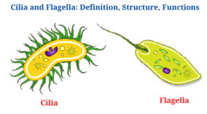
- An entire cell is propelled by its flagella (singular form: flagellum), which are long, hair-like projections that protrude from the plasma membrane.
- Small hairlike structures called cilia (singular = cilium) move entire cells (like paramecia) or substances along the cell’s outer surface (for example, the cilia of cells lining the Fallopian tubes that move the ovum toward the uterus, or cilia lining the cells of the respiratory tract that trap particulate matter and move it toward the nostrils).
- Arbitrarily, the terms cilium (which refers to an eyelash) and flagellum (which refers to a whip) are employed.
- It’s common knowledge that cilia are much smaller than flagella (as opposed to >40m).
- The number of cilia on the cell’s surface is substantially higher than on the inside (ciliated cells often have hundreds of cilia but flagellated cells usually have a single flagellum).
- However, the actual distinction can be found in the way the two move. Cilia are rowing like a crew on a boat. A biphasic movement occurs when the cilium is held rigid and only bends at the base of the cilium, followed by a recovery stroke in which the bend at the base extends to the tip.
- Flagella squirm around like a school of eels. In general, they generate waves that travel in a straight line from the bottom to the tip.
- In this way, water is moved in a flagellum in an orthogonal fashion to the flagellum’s axis, while water is moved perpendicularly in cilia along the cell’s surface.
Flagella and Cilia structure
Cilia as well as flagella are structurally indistinguishable, despite their differing patterns of beating.
All cilia as well as flagella have the same basic structure:
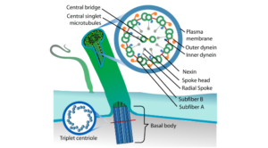
- The axoneme is a bundle of microtubules which is surrounded by a membrane which is part of the plasma membrane as well asis 1 to 2 nm in length as well as0.2 m in diameter.
- The axoneme is related to the basal body, which is an intracellular granule which starts from the centrioles as well asis found in the cell cortex.
- Each axoneme is full of ciliary matrix, which contains two inner singlet microtubules, each with 13 protofilaments, as well as nine outside doublet microtubules. The 9 + 2 array is the name for this recurrent motif.
- Each doublet has one full microtubule, referred known as the A sub fibre, which contains all 13 protofilaments. A B sub fibre with 10 protofilaments is attached to each A sub fibre.
- Subfibre A has two dynein arms which rotate clockwise. Nexin links are used to connect doublets.
- Dynein is an ATPase which transfers the mechanical labour of ciliary as well asflagellar beating into the energy generated by ATP hydrolysis.
- Radial spokes terminating in fork-like structures termed spoke knobs or heads connect each sub fibre A to the core microtubules.
Centrioles have a consistent arrangement of microtubules as well asrelated proteins with a nine-way pattern. Cilia as well asflagella, unlike centrioles, have a central pair of microtubules, resulting in the 9 + 2 axoneme configuration.
Note : which the composition of eukaryotic flagella differs from which of prokaryotes. Flagella in eukaryotes have a lot more proteins as well as look a lot like motile cilia, with similar motion as well as control patterns.
Cilia and Flagella Functions
In Eukaryotes
The dynein arms press on the adjoining outer doublets, causing them to slide together, using ATP produced by mitochondria near the base of the cilium or flagellum as fuel. Sliding is converted to bending because the arms are triggered in a strict sequence both around and down the axoneme, and the amount of sliding is restricted by the radial spokes and inter-doublet links.
In Prokaryotes
Bacterial flagella have a completely different mechanism than human flagella. The bacterial flagellum’s motion is fully controlled by the rotary motor at its base, similar to the propeller on a boat. The bacterial flagellum is a specialised extracellular cell wall component composed of only one protein (flagellin), which bears no resemblance to tubulin or dynein. Cilia as well as flagella are completely filled with cytosol up till their tips, as well as they use the ATP in which cytosol to create force along their whole length.
Cilia Functions
- Some protozoans and other single-celled organisms employ cilia for locomotion (e.g., Paramecium).
- The regular undulation of mobile cilia is used to effectively remove items such as dirt, dust, microorganisms, and mucus in order to keep people healthy.
- Cilia serve an important function in the cell cycle and in animal growth, including the development of the heart. Cilia are found throughout the body.
- A subset of proteins is enabled to function properly by cilia.
- Cia participate in cell communication and molecular trafficking as well, which is why they’re so important.
- A cell’s sensory equipment is provided by non-motile cilia, which picks up on signals. Sensory neurons depend on them for their functionality. Nephrons and retinal photoreceptors both have non-motile cilia for detecting urine flow.
- As a result, symbiotic microbiomes in animals have more places to live and reproduce.
- Ectosome vesicular secretion is aided by cilia, according to research.
Flagella Functions
- Flagella are utilised by cells such as the spermatozoon as well as Euglena to move about (protozoan).
- Flagella play an important function in eukaryotic reproduction as well as cell nutrition.
- Flagella serve as propelling motors in prokaryotes, such as bacteria, as well as are the primary means by which bacteria swim through fluids.
- It also gives pathogenic bacteria a way to help them colonise hosts as well as spread diseases.
- Flagella also serve as scaffolds or bridges for attachment to host tissue.
Cilia and Flagella Citations
- Stephen R. Bolsover, Elizabeth A. Shephard, Hugh A. White, Jeremy S. Hyams(2011). Cell Biology: A short Course (3 ed.).Hoboken,NJ: John Wiley and Sons.
- Alberts, B. (2004). Essential cell biology. New York, NY: Garland Science Pub.
- https://www.google.com/url sa=t&rct=j&q=&esrc=s&source=web&cd=3&cad=rja&uact=8&ved=2ahUKEwi3uLj09XeAhVYat4KHUh_D8sQFjACegQIBBAK&url=https%3A%2F%2Fcourses.lumenlearning.com%2Fboundless-biology%2Fchapter%2Fthe-cytoskeleton%2F&usg=AOvVaw3HEfpdUCrOziwpHgBB6XzG
- https://sciencing.com/main-functions-cilia-flagella-10572.html
Related Posts
- Phylum Porifera: Classification, Characteristics, Examples
- Dissecting Microscope (Stereo Microscope) Definition, Principle, Uses, Parts
- Epithelial Tissue Vs Connective Tissue: Definition, 16+ Differences, Examples
- 29+ Differences Between Arteries and Veins
- 31+ Differences Between DNA and RNA (DNA vs RNA)
- Eukaryotic Cells: Definition, Parts, Structure, Examples
- Centrifugal Force: Definition, Principle, Formula, Examples
- Asexual Vs Sexual Reproduction: Overview, 18+ Differences, Examples
- Glandular Epithelium: Location, Structure, Functions, Examples
- 25+ Differences between Invertebrates and Vertebrates
- Lineweaver–Burk Plot
- Cilia and Flagella: Definition, Structure, Functions and Diagram
- P-value: Definition, Formula, Table and Calculation
- Nucleosome Model of Chromosome
- Northern Blot: Overview, Principle, Procedure and Results

