Definition of Epithelial Tissue
One of the four types of tissues (muscular, epithelial, nervous, and connective) found inside animals is epithelial tissue. It is made up of tightly aggregated polyhedral cells which make cellular sheets which make a lining in the inside of vacant organs as well as cover up the body surface. An epithelial tissue, also called an epithelium (plural epithelia), is composed of cells organised in one or even more layers in continual sheets.
Characteristics of Epithelial Tissue
Regardless, the shape as well as purpose of epithelial tissue in diverse places of the body may be different, they all have some similarities.
The following are some of these characteristics:
- Shape and Size
- Epithelial cells appear in a range of forms as well as sizes, varying between towering columnar to cuboidal to low squamous.
- Appearance and dimension of cells are usually determined by their role.
- Polarity
- Organelles and membrane proteins are spread irregularly in epithelial cells, indicating polarity.
- The apical (free) surface of an epithelial cell confronts the skin, a body cavity, an internal organ lumen, or a gland duct that receives cell secretions. On apical surfaces, cilia or microvilli can be visible.
- Intercellular adhesion and other connections may be found on the lateral surfaces of epithelial cells that face neighbouring cells on each side.
- An epithelial cell’s basal surface clings to extracellular materials like the basement membrane, that is a still connective tissue generated by epithelial cells.
- Basement Membrane
- A slender extracellular layer composed of two layers: the basal and reticular lamina is the basement membrane.
- The basal lamina, which is nearer to as well as released by epithelial cells, includes proteins such as laminin and collagen, and specific glycoproteins and proteoglycans.
- The reticular lamina is nearer to the connective tissue below it, and collagen protein created by fibroblasts, connective tissue cells, is found there.
- Intercellular Adhesion and Other Junctions
- Adhesion and communication between cells are provided by a number of membrane-associated entities.
- Zonulae occludens, or tight junctions, seem to be the most popular apical of the junctions which entirely ring every cell.
- The adherens junction, also referred as the zonula adherens, envelops the epithelial cell and is easily noticeable underneath the tight junction.
- • The desmosome or macula adherens, which are disc-shaped structures, is another anchoring junction on one cell’s surface that match alike structures on the surface of an adjacent cell.
- Intercellular communication is mediated by gap junctions rather than cell adhesion or blockage.
- Avascular
- Epithelial tissue is avascular, depending upon the blood vessels of the fundamental connective tissue to give nutrients plus discharge waste.
- Diffusion allows chemicals to move between epithelium and connective tissue.
- Innervated
- Epithelial tissue is innervated. It means that Epithelial tissue has a nerve supply of its own.
7 . Renew and Repair
Epithelial cells divide quickly, permitting epithelial tissue to continuously replenish and heal itself by shedding damaged or diseased cells in addition to substituting them with new ones.
Functions of Epithelial Tissue
Epithelial tissue serves a variety of purposes depending on its location. Here are a few examples:
(a) Protection
- Amongst the utmost significant roles of epithelial tissue is protection. Desiccation, radiation, pathogen invasion, physical stress and toxins are all protected from the cells below.
- Since epithelial tissue does not have blood veins, bleeding inside the tissue following scratch is stopped.
(b) Transportation
- Several pumps available in epithelial tissue assist in the transfer of various substances into and out of cells.
- It also permits interchange of chemical substances among the fundamental cells as well as the ducts, capillaries, and body cavity in the respiratory, urine and digestive systems.
(c) Secretion
- Glandular epithelium releases a variety of macromolecules, including hormones, that are involved in a variety of biological processes.
- A lot of endocrine and exocrine glands too assist in the maintenance of body surfaces (skin) and the assisstance of a variety of organ functions (digestive system)
(d) Absorption
- Purpose of multiple specialised structures on the surface of cells, such as cilia and microvilli, helps in the assimilation of various molecules by expanding the surface area.
- Assimilation of water as well as other nutrients inside the digestive tract is aided by columnar cells inside the small intestine.
(e) Receptor function
- Some epithelial cells are specialised to execute sensory functions, allowing them to detect sensory data and transform it into brain signals.
- Apical cilia in epithelial tissue, such as the pseudostratified columnar epithelium of the olfactory mucosa, provide odour sensing.
Epithelial tissue Types / Classification with examples and location
Epithelial tissue is categorized into two kinds:
- The innermost layer of body cavities, ducts, blood vessels, as well as the digestive, respiratory, reproductive and urinar systems is the layering as well as coating epithelium, also known as the surface epithelium. It also serves as the exterior coating of blood arteries, ducts, and bodily cavities, and the outermost layer of the skin as well as several internal organs.
- The glandular epithelium formulates the emitting part of the glands like sweat glands, adrenal glands, digestive glands and thyroid gland.
- The structure of cells plus their shapes are used to classify different types of coating and covering epithelial tissue.
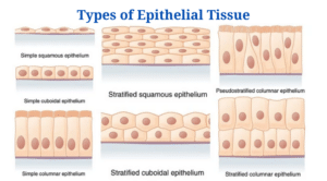
Simple epithelium
- On secretory and absorptive surfaces, simple epithelium is composed of only one layer of similar cells, that improves such functionalities.
- The three primary varieties of simple epithelium are named by the shapes of the cells, that varied depending on their roles.
(a) Simple squamous epithelium
- The simple squamous epithelium contains only one layer of flat cells which look like floor tiles when observed from the apical surface, with only a prominently positioned nucleus which is flattened and oval or spherical.
- Endothelium is the epithelial layer of serous membranes (peritoneum, pleura, pericardium) that covers the circulatory and lymphatic systems (heart, blood vessels, lymphatic vessels).
- Simple squamous epithelium is also in the air sacs of the lungs, the glomerular (Bowman’s) capsule of the kidneys, as well as the innermost side of the tympanic membrane (eardrum).
(b) Simple cuboidal epithelium
- Simple cuboidal epithelium is only one layer of spherical, cube-shaped cells with a nucleus inside the centre.
- It produces pigmented epithelium on the posterior aspect of the retina, coats kidney tubules and smaller ducts of several glands, and creates the secreting component of a number of glands like the thyroid gland plus ducts of certain glands like the pancreas.
(c) Simple columnar epithelium
- The columnar epithelium is composed of only one layer of rectangular cells on a basement membrane.
- This epithelium lines several organs as well as it is frequently developed to suit a particular purpose.
- The stomach is lined with columnar epithelium, which has no surface features. The free surface of the columnar epithelium lining the small intestine, on the other hand, is coated with microvilli, that give a large surface area for nutrient absorption.
- The columnar epithelium of the trachea is ciliated. It also contains mucus-secreting goblet cells, and eggs are pushed to the uterus by ciliary action in the uterine tubes.
Stratified Epithelium
- Basement membranes are frequently missing in a stratified epithelium, which is made up of many layers of cells of varied shapes.
- When basal cells divide, daughter cells arising through cell divisions push older cells upwards toward the apical layer; as they go more towards the surface and far from blood supply in underlying connective tissue, they get dehydrated and metabolically dormant.
- As the amount of cytoplasm in the cell decreases, tough proteins take over, as well as cells become harsh, hard structures that eventually die.
- The three main types of stratified epithelium are stratified squamous, stratified cuboidal, and stratified columnar epithelium.
- Dead cells are peeled off at the apical layer when they lose cell connections, but they are constantly replaced as new cells emerge from basal cells.
(a) Stratified squamous epithelium
The stratified squamous epithelium is comprised of two or even more layers of cells. The apical layer as well as many layers underneath it are made up of squamous cells, while the lower layers are made up of cuboidal to columnar cells.
1 . Keratinized squamous epithelium stratified
- In the apical section of cells, this epithelium forms a strong coating of keratin, which is many layers deep.
- • When cells travel away from nutrient-rich blood, the quantity of keratin inside cells increases, and organelles die.
- Keratin generates a strong, moderately impermeable defensive coating which keeps the living cells underneath from drying out.
- Keratinized stratified squamous epithelium forms a superficial layer of skin.
2. Non-keratinized stratified squamous epithelium
- This epithelium is hydrated continually by means of mucus from salivary as well as mucous glands and doesn’t include considerable levels of keratin inside the apical layer along with many layers deep.
- Wet surfaces (mouth, oesophagus, part of the epiglottis, part of the pharynx, and vagina) are lined with nonkeratinized stratified squamous epithelium, which also wraps the tongue.
(b) Stratified cuboidal epithelium
- The apical layer of stratified cuboidal epithelium is composed of cuboidal cells, whereas the lower layer might be either cuboidal or columnar.
- The excretory ducts of salivary and sweat glands have stratified cuboidal epithelium.
(c) Stratified columnar epithelium
- The stratified columnar epithelium is composed of numerous layers of cells, with columnar cells at apex as well as cuboidal or columnar cells in the deeper levels.
- The conjunctiva of the eyes, sections of the urethra, as well as a small area of the anal mucosa all have this type of epithelium.
Pseudostratified columnar epithelium
- The pseudostretified epithelium has somewhat multiple layers as the nuclei of the cells are present at a range of levels.
- Several cells don’t reach the apical surface while being connected to the basement membrane in only one layer.
- It looks to be a multilayered tissue as a result of these characteristics; however it is the basic epithelium, in reality.
- The epididymis, major ducts of a lot of glands, sections of the male urethra, and airways of the majority of the upper respiratory tract are all lined with this epithelium.
Transitional epithelium tissue
- The appearance of transitional epithelium (urothelium) varies (transitional).
- It resembles stratified cuboidal epithelium in an unstretched or comfortable form, with the exception that the apical layer cells are large and rounded.
- Cells become flatter as tissue is stretched, providing the look of stratified squamous epithelium. Its several layers along with suppleness make it suitable for lining hollow structures such as the urinary bladder, which are vulnerable to internal extension.
Glandular Epithelium
- Epithelial cells which primarily manufacture and secrete different macromolecules can be found in epithelia with other important roles or in glands, which are specialised organs.
- In simple columnar, simple cuboidal, and pseudostratified epithelia, scattered secretory cells, also known as unicellular glands, are frequent.
- From covering epithelia, glands emerge within the foetus by cell proliferation as well as extension in the fundamental connective tissue, then differentiation.
Endocrine Glands
- Hormones, which are secretions from endocrine glands, go into the interstitial fluid plus permeate further into bloodstream instead of passing through a duct.
- Because endocrine secretions are carried throughout the body by the bloodstream, they have far-reaching effects.
- Pituitary and pineal glands in the thyroid, parathyroid glands and in the brain close to the larynx (voice box), adrenal glands superior to kidneys, ovaries inside the pelvic cavity, pancreas inside the stomach, testes inside the scrotum, as well as thymus inside the thoracic cavity are all examples of endocrine glands.
Exocrine Glands
- Exocrine glands secrete one‘s substances into ducts, that ultimately discharge them on to skin’s surface or a hollow organ’s lumen.
- The impacts of exocrine gland secretions are restricted, but several of them can be hazardous when they enter the bloodstream.
- Sweat, oil, and earwax glands on the skin, and also digestive glands like salivary glands (secrete into the mouth cavity) and pancreas, are exocrine glands (secrete into the small intestine).
Epithelial Tissue: Types, Location and Functions
| Types of Epithelial Tissue | Location | Structure | Function |
| Simple squamous | Blood vessel lining, air sac lining of lungs | A single layer of flat cells having irregular boundaries | Transport by diffusion and where minimal protection is required |
| Simple Cuboidal | The tubular lining of kidneys, glandular ducts | A single layer of short cylindrical cells. It may have microvilli as in proximal convoluted tubules | Absorption and secretion |
| Simple Columnar | Digestive tract and upper respiratory tract lining | A single layer of columnar cells (tall and slender) and often ciliated | Protection, absorption, mucus secretion and movement in a specific direction |
| Stratified Squamous | The lining of the mouth and vagina | Made up of several layers of cells, continuously sloughed off and regenerated. The older layer of cells is pushed upwards and becomes flat. The lower layer is columnar and metabolically active | Protection |
| Stratified Cuboidal | Mammary glands, sweat gland and salivary glands | The upper layer is cuboid and other layers may be cuboidal or other types | Protection of ducts of various glands |
| Stratified Columnar | Male urethra and lobar ducts of salivary glands | There is a layer of columnar cells present on squamous, columnar or cuboidal epithelial cells | Protection and secretion |
| Pseudostratified Columnar | Respiratory passage and ducts of many glands | Similar to columnar epithelium but all the cells are not of similar height | Protection, secretion and movement of mucous |
| Transitional epithelia or urothelium | Urinary bladder, urethra, ureter | Stratified epithelium, which can contract or expand as per the requirement. Cells are cuboidal when not stretched, but when the organ stretches, then tissue gets compressed and cells appear irregular and squamous-shaped | Stretch readily to accommodate the different volume of liquids
Act as a barrier and have tight junctions to prevent reabsorption of toxic substances |
| Keratinised | The outer or apical layer of the cell | Mostly dead and devoid of nucleus and cytoplasm. The cytoplasm gets replaced by keratin, which makes the layer waterproof | Protection against abrasion |
Epithelial Tissue Citations
- Tortora GJ and Derrickson B (2017). Principles of Physiology and Anatomy. Fifteenth Edition. John Wiley & Sons, Inc.
- Waugh A and Grant A. (2004) Anatomy and Physiology. Ninth Edition. Churchill Livingstone.
- https://www.kenhub.com/en/library/anatomy/overview-and-types-of-epithelial-tissue
- https://quizlet.com/25768290/chapter-3-tissues-flash-cards/
- https://quizlet.com/47458913/chapter-4-flash-cards/
- https://quizlet.com/26692499/chapt-4-epithelial-tissue-flash-cards/
- https://quizlet.com/25669314/anatomy-ch-4-reading-flash-cards/
Related Posts
- Phylum Porifera: Classification, Characteristics, Examples
- Dissecting Microscope (Stereo Microscope) Definition, Principle, Uses, Parts
- Epithelial Tissue Vs Connective Tissue: Definition, 16+ Differences, Examples
- 29+ Differences Between Arteries and Veins
- 31+ Differences Between DNA and RNA (DNA vs RNA)
- Eukaryotic Cells: Definition, Parts, Structure, Examples
- Centrifugal Force: Definition, Principle, Formula, Examples
- Asexual Vs Sexual Reproduction: Overview, 18+ Differences, Examples
- Glandular Epithelium: Location, Structure, Functions, Examples
- 25+ Differences between Invertebrates and Vertebrates
- Lineweaver–Burk Plot
- Cilia and Flagella: Definition, Structure, Functions and Diagram
- P-value: Definition, Formula, Table and Calculation
- Nucleosome Model of Chromosome
- Northern Blot: Overview, Principle, Procedure and Results

