Opsonization Definition
- Opsonization means the ability of antibodies as well as complement components (and other proteins) to cover potentially harmful antigens, which can subsequently be detected by antibodies or complement receptors on phagocytic cells.
- Opsonization is The molecular process by which bacteria, apoptotic cells, or molecules are chemically changed to have greater connections with phagocyte as well as antibody cell surface receptors.
- It is the method by which intruding particles (antigens) are identified by the usage of particular components known as opsonins.
- Opsonins function as markers or tags which enable the body’s immune system to recognise them.
- An opsonin is some molecule which promotes phagocytosis by identifying an antigen for immune response or dead cells for recycling.
- Opsonization is used to create antigens more appealing to antibodies or phagocytic cells.
Opsonization Mechanism
Pathogens can be poisoned by antibodies or the complement system.
Antibody-mediated Opsonization
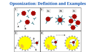
Figure: 1) Antibodies (A) and pathogens (B) free roam in the blood. 2) The antibodies bind to pathogens and can do so in different formations such as opsonization (2a), neutralization (2b), and agglutination (2c). 3) A phagocyte (C) approaches the pathogen, and the Fc region (D) of the antibody binds to one of the Fc receptors (E) on the phagocyte. 4) Phagocytosis occurs as the pathogen is ingested. Source: Wikipedia.
- Antibodies use the opsonization process to prevent and eliminate infection.
- Antibody-mediated opsonization by antibodies entails covering pathogens with antibodies in order for them to be identified as well as phagocytozed by inherent immune cells.
- When encapsulated bacteria which defy phagocytosis are covered with antibody, they appear to be exceedingly appealing to neutrophils and macrophages, and their rate of clearance from the circulation is dramatically increased.
- Like the structure of immunoglobulins was revealed, it became clear that IgG is the heat-stable serum component which helps in antigen-specific opsonization.
- IgG binds to antigenic determinants on the microorganism’s surface via its two antigen-binding components, Fab (or another particle).
- When IgG binds to antigen, it experiences conformational as well as configurational modifications in the F(ab)2 hinge region.
- All forms of phagocytic cells contain IgG receptors on their plasma membranes.
- Amount of such receptors on every mouse peritoneal as well as alveolar membrane has indeed been reported to be between 1-2 million. Such receptors withstand tryptic proteolysis and mediate IgG-coated particle binding at 4°C, 37°C, and in the nonexistence of divalent cations.
- Despite the fact that all four subclasses of human IgG attach to the antigen, only IgG1 and IgG3 can connect to receptors on phagocytic cells.
- IgG receptors on phagocytic cells only attach to the Fc region of the molecule and are so classified as Fc receptors. Attachment of a pathogen (antigen)-antibody complex to an Fc receptor on phagocytes triggers internalisation of the complex as well as inner digestion of the pathogen in lysosomes in the case of antibody-mediated opsonization.
- Several antibodies, in fact, attach to different places on the antigen, enhancing the likelihood plus effectiveness with which the pathogen is swallowed in the phagosome and killed by lysosomes.
Complement-mediated Opsonization
- Opsonization complement system is made up of around 30 proteins which help antibodies as well as phagocytic cells fight off intruding organisms.
- Through opsonizing antigens, it promotes phagocytosis. This mechanism is also in charge of promoting inflammation as well as cytolysis.
- C3b is by far very crucial heat-labile opsonin, and maybe very important opsonin of all (C3b is the fragment of C3 which connects to particles when C3 is cleaved by a C3-convertase).
- C3 is cleaved into C3a and C3b via the standard pathway (started by attachment of IgG or IgM molecules to antigen, that results in attachment and stimulation of the C 1 complex) or the alternative approach (started by the existence of lipid-carbohydrate complexes present within cell wall of bacteria).
- C3b attaches to the surface of the particle and acts as an opsonin.
- Until a particle may be ingested, it ought to first be identified by and bonded to the surface of a phagocytic cell.
- The processes that cause these events have indeed been partly identified. Every mononuclear phagocytes as well as polymorphonuclear leukocytes examined so far contain C3b receptors on their plasma membranes.
- The attachment of C3b-coated particles to C3b receptors in certain cells, on the other hand, involves the existence of divalent cations in the medium.
- Activated macrophages subsequently consume C3b-coated particles.
- Furthermore, because C3b has an enzymatic activity for aromatic dipeptides, it cleaves the aromatic dipeptides contained in neutrophils in bacteria such as Hemophilus influenza.
- Cleavage of aromatic dipeptides on the plasma membrane of neutrophils is thought to be a mechanism through which C3b facilitates phagocytosis of particles to which it is attached.
Opsonins and Types
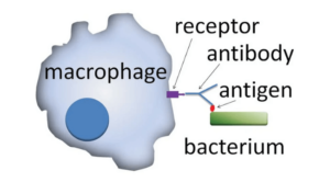
Figure: Action of opsonins: A phagocytic cell recognizes the opsonin on the surface of an antigen. Source: Wikipedia.
- Every molecule that facilitates phagocytosis by recognising an antigen for immune response or dead cells for recycling is an opsonin.
- Opsonin molecules typically attach to the receptors found in the antigen on one end and the receptors on the phagocytes on the other.
- This binding results in many mechanisms that eventually lead to the degradation or elimination of the specific antigen.
- Opsonin molecules guarantee that antigen attaching to immune cells is considerably improved.
- Opsonins play critical functions in the immune system, such as identifying dead and dying cells for clearance by macrophages and neutrophils.
- Opsonins also assist in the activation of complement proteins and the killing of cells by natural killer (NK) cells.
- Opsonins use methods to improve the speed of phagocytosis by promoting the communication among opsonin and cell surface receptors on immune cells.
- Such chemicals overcome the negative charges on the cell membrane, making it harder for two cells to come into contact.
Types
- Opsonins occupied in the immune system comprise the compounds listed below:
Antibodies
- Adaptive immunity molecules which are discharged by B cells in response to an immune response are antibodies.
- In case of IgG antibodies, the occurrence of the Fc domain permits receptors on phagocytes to connect to the Fc domain, whereas the antibody’s Fab domain connects to the antigen.
- IgM antibodies, on the other hand, lack Fc receptors and so are useless in increasing phagocytosis. However, they are quite efficient at starting the complement system and are classified as an opsonin.
- The binding of antibodies to antigens and immune cells causes the effector cells to produce lysis products.
Complement Proteins
- C3b, C4b, and C1q are the most prevalent complement proteins which also function as opsonins.
- Because it can be identified by phagocyte receptors, C3b is a very efficient opsonin which stimulates phagocytosis.
- Complement proteins acting as opsonin have the added benefit of being expressed in all phagocytes and recognising many complement proteins such as C3b and C4b.
- C1q is a component of the C1 complex that works as an opsonin by interacting with the Fc region of antibodies.
Circulating Proteins
- A variety of circulating proteins, including pentraxins, collectins, and ficolins, function as opsonins.
- Such proteins are PRRs (Pattern Recognising Receptors), which can bind microorganisms as an opsonin and increase neutrophil activity through a variety of methods.
- The ability of these proteins to attach in a Cadependent form, as a pattern recognition molecule, to various bacteria which have phosphorylcholine in their membranes, which then triggers the complement system, is a noteworthy trait.
- The lectin route of complement activation is founded on collectins such as Mannose Binding Lectin (MBL).
- Ficolins, in turn, often recognise Nacetylglucosamine residues in complextype carbohydrates, as well as other ligands on various antigens.
Examples
The following are examples of common opsonins:
Immunoglobulin M (IgM) antibodies
- Anti-IgG antibodies
- Proteins of the C3b class
- Proteins of the C4b family
- C1q proteins are a type of protein that is found in the
- Pentraxins \sCollectins
- Ficolins
- lectin that binds to mannose (MBL)
Opsonization Citations
- Thau L, Mahajan K. Physiology, Opsonization. [Updated 2020 Mar 25]. In: StatPearls [Internet]. Treasure Island (FL): StatPearls Publishing; 2020 Jan-. Available from: https://www.ncbi.nlm.nih.gov/books/NBK534215/
- Griffin F.M. (1977) Opsonization. In: Day N.K., Good R.A. (eds) Biological Amplification Systems in Immunology. Comprehensive Immunology, vol 2. Springer, Boston, MA
- https://www.ncbi.nlm.nih.gov/books/NBK534215/
- https://patents.google.com/patent/US20040013673A1/en
- https://makkypedia.blogspot.com/2016/06/opsonization.html
- https://www.sciencedirect.com/topics/immunology-and-microbiology/immunoglobulin-g
- https://www.sciencedirect.com/book/9780120442201/methods-for-studying-mononuclear-phagocytes
- https://www.ncbi.nlm.nih.gov/pmc/articles/PMC4139653/
Related Posts
- Phylum Porifera: Classification, Characteristics, Examples
- Dissecting Microscope (Stereo Microscope) Definition, Principle, Uses, Parts
- Epithelial Tissue Vs Connective Tissue: Definition, 16+ Differences, Examples
- 29+ Differences Between Arteries and Veins
- 31+ Differences Between DNA and RNA (DNA vs RNA)
- Eukaryotic Cells: Definition, Parts, Structure, Examples
- Centrifugal Force: Definition, Principle, Formula, Examples
- Asexual Vs Sexual Reproduction: Overview, 18+ Differences, Examples
- Glandular Epithelium: Location, Structure, Functions, Examples
- 25+ Differences between Invertebrates and Vertebrates
- Lineweaver–Burk Plot
- Cilia and Flagella: Definition, Structure, Functions and Diagram
- P-value: Definition, Formula, Table and Calculation
- Nucleosome Model of Chromosome
- Northern Blot: Overview, Principle, Procedure and Results

