The Golgi apparatus has multiple names such as Golgi complex or Golgi body. The name is given on the name of the scientist, who discovered the organelle, i.e. Camillo Golgi. It is found in all the eukaryotic cells, plants as well as animals. They are membrane-bound organelle present in the cytosol of the cell. Let us discuss more about Golgi complex.
Golgi Apparatus Definition
- The Golgi apparatus, also known as the Golgi body, Golgi complex, or simply Golgi, is a cellular organelle found in most eukaryotic cells.
- The cell’s manufacturing and shipping centre is referred to as this.
- Before protein molecules are delivered to their destination, Golgi is engaged in their packing. These organelles assist in the processing and packaging of macromolecules created by the cell, such as proteins and lipids, and so serve as the cell’s “post office.”
- Camillo Golgi, an Italian scientist, developed the Golgi apparatus in 1898.
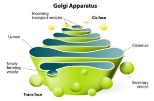
Golgi Apparatus Diagram
Golgi Apparatus Structure
- The Golgi apparatus appears to be made up of stacks of flattened structures containing many vesicles harbouring secretory granules under the electron microscope.
- In both plant and animal cells, the Golgi apparatus has a remarkably similar morphology. It is, however, exceedingly pleomorphic, appearing compact and restricted in some cell types and stretched out and reticular in others (net-like).
- The Golgi apparatus, on the other hand, is typically seen as a complicated network of interconnected tubules, vesicles, and cisternae.
A. Cisternae
- The cisterna is the basic unit of the Golgi apparatus.
- Cisternae are central, flattened, plate-like or saucer-like closed compartments that are held in parallel bundles or stacks one above the other and have a diameter of about 1 m.
- Cisternae are separated by a 20 to 30 nm interval in each stack, which may contain rod-like components or fibres.
- In animal cells, each stack of cisternae creates a dictyosome, which may include 5 to 6 Golgi cisternae or 20 or more in plant cells.
- A smooth unit membrane (7.5 nm thick) surrounds each cisterna, with a lumen ranging in width from 500 to 1000 nm.
- Each cisterna’s edges are softly curved, giving the Golgi apparatus’s complete dictyosome a bow-like look.
- The proximal, forming or cis-face cisternae are found near the convex end of the dictyosome, whereas the distal, maturing or trans-face cisternae are found at the concave end.
B. Tubules
- The dictyosome is surrounded by and radiates from a complex array of related vesicles and anastomosing tubules (30 to 50 nm diameter). In reality, the dictyosome’s periphery is fenestrated (lace-like) in structure.
C. Vesicles
There are three types of vesicles (60 nm in diameter):
- Transitional vesicles are small membrane-limited vesicles that migrate and converge to the cis face of the Golgi, where they merge to create new cisternae, and are hypothesised to form as blebs from the transitional ER.
- Secretory vesicles are membrane-bound vesicles that discharge from the Golgi cisternae’s edges. They frequently arise between the Golgi maturing face and the plasma membrane.
- Clathrin-coated vesicles are spherical protuberances with a rough surface and a diameter of around 50 m. They are physically distinct from secretory vesicles and are present near the organelle’s perimeter, usually at the extremities of solitary tubules. Clathrin-coated vesicles are known to have a function in membrane and secretory product intracellular trafficking, such as between the ER and the Golgi, as well as between the GELR area and the endosomal and lysosomal compartments.
How to increase Brain Power – Secrets of Brain Unlocked
Golgi Apparatus Functions
1 . Golgi vesicles are commonly referred to as the cell’s “traffic cops.” They play an important role in sorting and guiding many of the cell’s proteins and membrane components to their right destinations.
- To carry out this function, Golgi vesicles contain different sets of enzymes in different types of vesicles—cis, middle, and trans cisternae—that react with and modify secretory proteins passing through the Golgi lumen, as well as membrane proteins and glycoproteins that are transiently in the Golgi membranes on their way to their final destinations.
- As a result, the Golgi apparatus serves as the cell’s assembly factory, with raw materials being sent to it before being passed out of the cell.
2. The Golgi apparatus is responsible for the packing and exocytosis of the following materials in animals:
- Exocrine pancreatic cell zymogen;
- Mucus (=a glycoprotein) production by intestinal goblet cells;
- Mammary gland cells secrete lactoprotein (casein) (Merocrine secretion);
- Thyroid cells secrete thyroxine hormone molecules (thyroglobulins);
- Tropocollagen and collagen secretion
- Melanin granules and other pigments are formed; and
- The yolk and vitelline membranes of expanding primary oocytes are formed.
3. It also plays a role in the creation of cellular organelles such the plasma membrane, lysosomes, spermatozoa acrosomes, and oocyte cortical granules.
4. They also play a role in lipid molecule trafficking throughout the cell.
5. In addition, the Golgi complex is involved in the formation of proteoglycans. Proteoglycans are a type of chemical found in the extracellular matrix of animal cells.
6. It is also an important location for carbohydrate production. Glycosaminoglycans are synthesised, and Golgi attaches to these polysaccharides, which then attach to a protein generated in the endoplasmic reticulum to form proteoglycans.
7. The Golgi has a role in the sulfation of certain compounds.
8. The import of ATP into the Golgi lumen is required for the phosphorylation of molecules by the Golgi.
9. In plants, the Golgi apparatus is primarily responsible for the secretion of primary and secondary cell wall components (e.g., formation and export of glycoproteins, lipids, pectins and monomers for hemicellulose, cellulose, lignin, etc.)
Click Here for Complete Biology Notes
Golgi Apparatus Citations
- Stephen R. Bolsover, Elizabeth A. Shephard, Hugh A. White, Jeremy S. Hyams(2011). Cell Biology: A short Course (3 ed.).Hoboken,NJ: John Wiley and Sons.
- Alberts, B. (2004). Essential cell biology. New York, NY: Garland Science Pub.
- https://biology.tutorvista.com/animal-and-plant-cells/golgi-apparatus.html
- http://www.biologydiscussion.com/cell/golgi-complex/golgi-complex-structure-and-functions-with-diagram/36799
Related Posts
- Phylum Porifera: Classification, Characteristics, Examples
- Dissecting Microscope (Stereo Microscope) Definition, Principle, Uses, Parts
- Epithelial Tissue Vs Connective Tissue: Definition, 16+ Differences, Examples
- 29+ Differences Between Arteries and Veins
- 31+ Differences Between DNA and RNA (DNA vs RNA)
- Eukaryotic Cells: Definition, Parts, Structure, Examples
- Centrifugal Force: Definition, Principle, Formula, Examples
- Asexual Vs Sexual Reproduction: Overview, 18+ Differences, Examples
- Glandular Epithelium: Location, Structure, Functions, Examples
- 25+ Differences between Invertebrates and Vertebrates
- Lineweaver–Burk Plot
- Cilia and Flagella: Definition, Structure, Functions and Diagram
- P-value: Definition, Formula, Table and Calculation
- Nucleosome Model of Chromosome
- Northern Blot: Overview, Principle, Procedure and Results

