Meiosis Definition
- In sexually reproducing eukaryotes, meiosis is a type of cell division that produces four daughter cells (gametes), each with half the number of chromosomes as the diploid parent cell.
- The haploid cells become gametes, which determine sexual reproduction and the birth of a new generation of diploid creatures by fusing with another haploid cell during fertilisation.
- Meiosis is a process that occurs in sexually reproducing organisms’ germ cells.
- Germ cells are found in the gonads of both plants and animals, although the timing of meiosis varies depending on the organism.
What is the Purpose of Meiosis ?
For the following reasons, meiosis is required for all sexually reproducing organisms:
- Through the generation of gametes, meiosis keeps the number of chromosomes in sexually reproducing organisms constant.
- Meiosis creates genetic variety among species by causing the exchange of genes that occurs when cells cross over. These differences are the building blocks of the evolutionary process.
Meiosis Stages and Phases
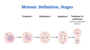
- Meiosis is made up of two stages of cell division: Meiosis I and Meiosis II.
- A period of karyokinesis (nuclear division) and cytokinesis is included in each round of division (cytoplasmic division).
Meiosis I
- The first meiotic division is characterised by a lengthy prophase during which homologous chromosomes come into close proximity to one another and exchange genetic material.
- Similarly, during the initial meiotic division, the number of chromosomes is reduced, resulting in the formation of two haploid cells.
- The heterotypic division is another name for the first meiotic division.
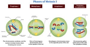
Figure: Phases of Meiosis I. Image Source: Wikipedia (Ali Zifan).
The steps of Meiosis I are as follows:
Interphase
- Meiosis, like mitosis, includes a preliminary phase known as interphase.
- The following characteristics define the interphase:
- The nuclear membrane is unaffected, and the chromosomes appear as scattered, lengthy, coiled, and barely visible chromatin fibres.
- The amount of DNA doubles. The size of the nucleolus is greatly enhanced due to the accumulation of ribosomal RNA (rRNA) and ribosomal proteins in the nucleolus.
- An interphase cell has two pairs of centrioles because a daughter pair of centrioles forms near the already existing centriole in animal cells.
- There is a significant alteration in the G2 phase of interphase that drives the cell toward meiosis rather than mitosis.
- The nucleus of the dividing cell begins to expand in size by taking water from the cytoplasm at the start of the first meiotic division, and the nuclear volume grows threefold.
Prophase I
- The longest stage of meiotic division is prophase I. It is divided into the following stages:
Leptotene
- The chromosomes grow even more uncoiled and resemble a long thread-like shape in the leptotene stage, and they develop chromomeres, which are bead-like structures.
- Because the chromosomes in the nucleus are still oriented towards centrioles at this stage, the nucleus looks like a bouquet in the animal cell. As a result, this stage is sometimes referred to as the Bouquet Stage.
Synaptotene vs. Zygotene
- The zygotene stage begins with synapsis, or the pairing of homologous chromosomes.
- A protein-containing structure called a synaptonemal complex connects the paired homologous chromosomes.
- The synaptonemal complex aids in the stabilisation of homologous chromosomal pairing and the facilitation of recombination and cross-over.
- The synapsis could start at one or more sites along the homologous chromosomes’ length.
- Synapsis can begin at the ends of chromosomes and progress to their centromeres (proterminal synapsis), or it can begin at the centromere and progress to the ends (intermediate synapsis) (procentric pairing).
- Synapsis can occur at various places on homologous chromosomes in some situations (random pairing).
Pachytene
- The chromosomes become coiled spirally around each other at this point, and they can no longer be recognised.
- Each homologous chromosome separates lengthwise to generate two chromatids in the midst of the pachytene stage, but they remain linked by their common centromere.
- Because there are two visible chromosomes at this time, the chromosomes are referred to as bivalent, or tetrad, because there are four visible chromatids.
- This stage is particularly important since it is during this stage that a significant genetic phenomena known as “crossing over” occurs.
- Redistribution and mutual exchange of genetic material between two homologous chromosomes are involved in crossing over.
- Endonuclease is an enzyme that splits non-sister chromatids at the point where they cross across.
- Following the breakage of chromatids, chromatid segments are exchanged between non-sister chromatids of homologous chromosomes.
- Ligase, a different enzyme, joins the broken chromatid segments to the non-sister chromatid.
- The crossing over process is the mutual exchange of chromatin material between one non-sister chromatid of each homologous chromosome.
Diplotene
- The synaptonemal complex appears to have disintegrated, leaving the chromatids of the paired homologous chromosome physically linked at one or more localised sites known as synaptonemal junctions.
- Chiasmata travel in a zip-like pattern towards the end of chromosomes in diplotene.
Diakinesis
- The bivalent chromosomes in the nucleus become more compacted and uniformly distributed at this stage.
- The nuclear envelope disintegrates at this point, and the nucleolus vanishes.
- The chiasmata also reach the chromosomal ends, and the chromatids remain joined until metaphase.
Metaphase I
- Spindle fibre attachment to chromosomes and chromosomal alignment near the equator characterise Metaphase I.
- The spindle fibres are connected to the homologous chromosomes’ centromeres, which are oriented towards the opposing poles, during metaphase I.
Anaphase I
- At anaphase I, homologous chromosomes are split from one another, and each homologous chromosome, with its two chromatids and undivided centromere, moves towards the opposite poles of the cell due to the shortening of chromosomal fibres or microtubules.
- Because one of the chromatids changed its counterpart during the chiasma development, the two chromatids of a chromosome are not genetically identical.
Telophase I
- The migration of a haploid set of chromosomes at each pole marks the start of telophase I.
- The chromosomes become uncoiled as the nuclear membrane forms around them. The nucleolus returns, resulting in the formation of two daughter nuclei.
Cytokinesis I
- In animals, cytokinesis happens when the cell membrane constricts, whereas in plants, it occurs when the cell plate forms, resulting in the development of two daughter cells.
Meiosis II
- The haploid cell splits mitotically in the second phase of meiotic division, resulting in four haploid cells. The homotypic division is another name for this division.
- As with the first meiotic division, this division does not contain the exchange of genetic material or the reduction of chromosomal number.
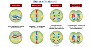
Figure: Phases of Meiosis II. Image Source: Wikipedia (Ali Zifan).
The steps of Meiosis II are as follows:
Prophase II
- Each centriole divides in prophase II, resulting in two pairs of centrioles.
- The nucleolus vanishes when the centrioles travel towards the opposing poles and the nuclear membrane.
Metaphase II
- The chromosomes are positioned on the equator of the cell by the spindle fibres during metaphase II.
- Each chromosome creates two daughter chromosomes when the centromere splits.
- Each chromosome’s centromere is connected to the spindle apparatus.
Anaphase II
- Due to the stretching of interzonal microtubules of the spindle and the shortening of chromosomal microtubules, the daughter chromosomes travel towards the opposing poles.
Telophase II
- Chromosomes are formed when chromatids move to opposite poles.
- The nucleolus reappears due to the synthesis of ribosomal RNA, and the endoplasmic reticulum forms the nuclear envelope around the chromosomes.
Cytokinesis II
- The cytokinesis process is identical to cytokinesis I, culminating in cytoplasm division for each of the four daughter cells.
Meiosis Applications
Meiosis has a variety of applications
Several lab-based technologies use meiotic like mitosis, some of which are listed below:
Tissue culture
- Meiosis, like mitosis, is employed in biotechnology to get cells into a gametic state.
- Meiosis frequently occurs in conjunction with mitosis to generate variety, which contributes in the study of evolutionary processes.
Gamete formation in vitro
- Embryonic stem cells are developed into germ-like cells through meiotic division in various gamete failure-related infertility disorders.
- These gametes are created in vitro by meiosis and injected into people who have genetic abnormalities.
Click Here for Complete Biology Notes
Meiosis Citations
- Rastogi SC (2006). Cell and Molecular Biology. Second Edition. New Age International.
- Eguizabal C, Montserrat N, Vassena R, et al. Complete meiosis from human induced pluripotent stem cells. Stem Cells. 2011;29(8):1186‐ DOI:10.1002/stem.672
- Ronchi VN (1995). Eguizabal C, Montserrat N, Vassena R, et al. Complete meiosis from human induced pluripotent stem cells. Stem Cells. 2011;29(8):1186‐ DOI:10.1002/stem.672.
- https://www.biologydiscussion.com/invertebrate-zoology/meiotic-division-of-cell-with-diagram/28053
- https://studywrap.com/meiosis-reproductive-cell-division-and-its-significance/
- https://www.coursehero.com/file/p7mfmf11/proposed-in-1953-by-Alma-Howard-and-Stephen-Pelc-of-Hammersmith-Hospital-London/
- http://www.notesonzoology.com/cytology/meiosis-and-cell-division-cytology/2221
- https://www.coursehero.com/file/p5s9706/f-The-nucleolus-and-nuclear-membrane-reappear-and-thus-two-daughter-nuclei-are/
- https://www.coursehero.com/file/p5ddfsk/Meiosis-consists-of-two-rounds-of-cell-division-Meiosis-I-and-Meiosis-II-Each/
- https://www.biologyexams4u.com/2012/09/crossing-over-process-behind-our.html
- https://quizlet.com/109271499/chapter-two-flash-cards/
- https://en.mimi.hu/biology/meiosis.html
- https://edurev.in/studytube/Meiosis-Stages-Cell-Cycle-and-Cell-Division–Biolo/4433ebb1-1a3c-449b-a33d-b7408c82bd93_t
- https://www.thoughtco.com/stages-of-meiosis-373512
- https://www.sciencedirect.com/topics/biochemistry-genetics-and-molecular-biology/meiosis
- https://www.sciencedirect.com/topics/biochemistry-genetics-and-molecular-biology/nucleolus
- https://www.researchgate.net/publication/309308410_Alignment_of_Homologous_Chromosomes_and_Effective_Repair_of_Programmed_DNA_Double-Strand_Breaks_during_Mouse_Meiosis_Require_the_Minichromosome_Maintenance_Domain_Containing_2_MCMDC2_Protein
Related Posts
- Phylum Porifera: Classification, Characteristics, Examples
- Dissecting Microscope (Stereo Microscope) Definition, Principle, Uses, Parts
- Epithelial Tissue Vs Connective Tissue: Definition, 16+ Differences, Examples
- 29+ Differences Between Arteries and Veins
- 31+ Differences Between DNA and RNA (DNA vs RNA)
- Eukaryotic Cells: Definition, Parts, Structure, Examples
- Centrifugal Force: Definition, Principle, Formula, Examples
- Asexual Vs Sexual Reproduction: Overview, 18+ Differences, Examples
- Glandular Epithelium: Location, Structure, Functions, Examples
- 25+ Differences between Invertebrates and Vertebrates
- Lineweaver–Burk Plot
- Cilia and Flagella: Definition, Structure, Functions and Diagram
- P-value: Definition, Formula, Table and Calculation
- Nucleosome Model of Chromosome
- Northern Blot: Overview, Principle, Procedure and Results

