Transitional Epithelium Definition
Transitional epithelium is a sort of stratified epithelium made up of numerous layers of cells whose shape changes depending on the organ’s function. When the epithelium is relaxed, it appears cubical or round, with the exception of the apical layer, which appears flattened when stretched. Because this epithelium is almost exclusively found in the urinary system, it is sometimes known as “urothelium.”
Structure of Transitional Epithelium
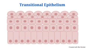
- Transitional epithelium is a type of epithelial tissue that looks as a stratified cuboidal epithelium when relaxed.
- The transitional epithelium’s cells are pear-shaped or spherical, but when the tissue stretches, the cells flatten out, producing the appearance of stratified squamous epithelium.
- The cells in the basal layer seem cuboidal or columnar, whereas the cells in the apical layer flatten as the degree of extension increases.
- The epithelium’s cell layers are classified into three types.
- The basal layer, which is immediately linked to the basement membrane, is the lowest layer. This membrane is in charge of supplying the top layers with continual cell renewal.
- The cytoplasmic proteins in the cells of the basal layer bind to create tonofibrils, which then converge with hemidesmosomes to form an effective link with the basement membrane.
- The cells in the basal layer have a lot of mitochondria because they need a lot of energy to regenerate the epithelium.
- The intermediate layer, or middle layer, is made up of highly proliferative, fast dividing cells that offer rapid regeneration when existing cells are injured or damaged.
- The Golgi apparatus, which facilitates in the transport of proteins like keratin to the superficial layers, is prevalent in this layer.
- The superficial layer’s cells are strongly keratinized, creating a barrier against salts and water.
- The superficial layer, also known as the apical layer, borders the lumen and shields the underlying layer of cells from pathogens in the lumen.
- Some of the cells in the superficial layer have microvilli and are mucus-coated.
- Gap junctions and desmosomes join the cells in the epithelium. The epithelium can expand because of these structural features, but the cells become brittle as a result.
- The transitional epithelium, like all other epithelial tissues, is avascular, meaning it lacks blood vessels. For oxygen, nutrition, and excretion, the cells of this epithelium rely on the blood vessels of the neighbouring connective tissues.
- The cells, on the other hand, have their own nerve supply
Transitional Epithelium Functions
The transitional epithelium has two major functions, which are determined by the cell’s structure and composition:
1. Permeability barrier
- The tissue provides exceptional impermeability to water and other molecules due to the presence of significant concentrations of keratin in the cells.
- The tissue’s cells are extremely resistant to osmotic pressure, preventing desiccation even when the cells are fully stretched.
- The re-entry of toxins and substances into the bloodstream is also prevented.
- The urinary system is a famous example of this function, as the cells in the urothelium are not dehydrated even when hypertonic urine is present in the lumen.
2 . Volume control
- The ability of this epithelium to stretch and modify the shape of the organs as the fluid pressure increases is the epithelium’s second crucial role.
- When the volume of fluid in the urinary bladder and ureters rises, the cells in the superficial layer of the excretory system stretch, changing their form from round to flat.
- Stretching the organs increases their volume while also protecting the underlying tissue from the toxins in the urine.
Transitional Epithelium Location and Examples
- The urothelium is the most well-known transitional epithelium.
- The transitional epithelium, also known as the urothelium, lines the urine bladder, ureters, and portions of the urethra.
- The transitional epithelium, which is continuous with the urothelium of the urine bladder, also lines the lining of the prostatic urethra in the male reproductive system.
Click Here for Complete Biology Notes
PhD in Psychology : Career, Admission Process, Benefits, Opportunities.
Transitional Epithelium Citations
- https://biologydictionary.net/transitional-epithelium/
- https://www.sciencedirect.com/science/article/pii/B978012802926800001X
- https://www.sciencedirect.com/science/article/pii/0002939461919511
- https://www.researchgate.net/publication/235443030_BLOOD-TISSUE_BARRIERS_Morphofunctional_and_Immunological_Aspects_of_the_Blood-Testis_and_Blood-Epididymal_Barriers
- https://www.quia.com/jg/838874list.html
- https://training.seer.cancer.gov/anatomy/urinary/
- https://quizlet.com/14186293/anatomy-chapter5-flash-cards/
- https://en.wikipedia.org/wiki/Keratinocyte
- https://www.ncbi.nlm.nih.gov/pmc/articles/PMC3579394/
Related Posts
- Phylum Porifera: Classification, Characteristics, Examples
- Dissecting Microscope (Stereo Microscope) Definition, Principle, Uses, Parts
- Epithelial Tissue Vs Connective Tissue: Definition, 16+ Differences, Examples
- 29+ Differences Between Arteries and Veins
- 31+ Differences Between DNA and RNA (DNA vs RNA)
- Eukaryotic Cells: Definition, Parts, Structure, Examples
- Centrifugal Force: Definition, Principle, Formula, Examples
- Asexual Vs Sexual Reproduction: Overview, 18+ Differences, Examples
- Glandular Epithelium: Location, Structure, Functions, Examples
- 25+ Differences between Invertebrates and Vertebrates
- Lineweaver–Burk Plot
- Cilia and Flagella: Definition, Structure, Functions and Diagram
- P-value: Definition, Formula, Table and Calculation
- Nucleosome Model of Chromosome
- Northern Blot: Overview, Principle, Procedure and Results









the content is just on point
thank you so much