What is a Cell?
Cells are the basic building blocks of all living things. The human body is composed of trillions of cells. They provide structure for the body, take in nutrients from food, convert those nutrients into energy, and carry out specialized functions. Cells also contain the body’s hereditary material and can make copies of themselves.
All organisms are made up of cells. They may be made up of a single cell (unicellular), or many cells (multicellular). Mycoplasmas are the smallest known cells. Cells are the building blocks of all living beings. They provide structure to the body and convert the nutrients taken from the food into energy.
Cells are complex and their components perform various functions in an organism. They are of different shapes and sizes, pretty much like bricks of the buildings. Our body is made up of cells of different shapes and sizes.
Discovery of living cell
By Anton Von Leuwenhoek
Discovery of cells is one of the remarkable advancements in the field of science. It helps us know that all the organisms are made up of cells, and these cells help in carrying out various life processes. The structure and functions of cells helped us to understand life in a better way.
Discovery of Nucleus –
By Robert Brown
Cell theory-
- Matthias Schleiden, a German Botanist, examined that plants & animals are composed of a cell.
- Theodore Schwann, a British Zoologist, reported the presence of plasma membrane.
- This didn’t explain how new cells are formed.
- Rudolf Virchow explained cells divided and new cells are formed from preexisting cells (Omnis cellula e cellula)
About cell-
- The cytoplasm is the main arena of cellular activities.
- Organelles – Membrane-bound distinct structures.
- Ribosomes – Cytoplasm, chloroplast, mitochondria, E.R.
- Animal – Centrosome (help in cell division)
- Smallest cell – Mycoplasma (0.3 micrometers)
- Largest isolated cell – Egg of ostrich
- Tumor RBC – 7 micrometers in diameter
- The largest cell in the human is – Nerve cell.
Type of Cell
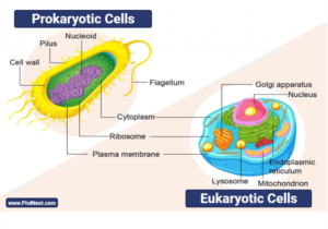
Cells may be typified in different ways. For instance, based on the presence of a well-defined nucleus, a cell may be eukaryotic or prokaryotic. Cells may also be classified based on the number of cells that make up an organism, i.e. ”unicellular”, ”multicellular”, or ”acellular
(a) PROKARYOTIC CELL
- E.g., Bacteria, Blue-green algae, mycoplasma
- Divide more rapidly.
- All have cell walls except mycoplasma.
- No nuclear membrane, naked genetic material.
- Linear and Circular DNA(Plasmid).
- Resistant to antibodies.
- Have mesosomes (infoldings of the cell membrane). E.g., Tubules (tube-like), vesicle(spherical), lamellae (highly folded).
- Mesosomes help in cell wall formation, DNA replication, respiration.
- Difference densities of nucleoplasm & cytoplasm prevent mixing of the Two.
- Cyanobacteria – Membranous Structure in the cytoplasm – chromatophores (contain pigment).
Cell Envelope-
- Outermost – glycocalyx, cell wall, plasma membrane.
- Glycocalyx as loose sheath – Slime layer (prevent dryness) as thick and tough – capsule
- Prevent cells from bursting or collapsing.
On the basis of staining – (By crystal violet)
Gram-positive – can be stained; Gram-negative – can’t be stained.
Motile or non-motile –
- If motile – filamentous extension called flagella.
- Bacterium flagellum – filament, hook, and the basal body.
- Filament – Largest portion; (whiplash or tinsel)
- Pili – Elongated tubular Structure but don’t play a role in motility.
- Fiber – Small bristle-like fibers (on the surface).
- They attach bacteria to rocks in the stream and host tissues.
Ribosomes and Inclusion bodies –
- Form helical Structure coiled together.
- Prokaryotes – !5nm-20nm – 50 s+30s = 70s
- Attach to single mRNA and form chain – polyribosome/polysome; they translate mRNA (messenger RNA) – protein; t- RNA- (transfer); rRNA(Ribosomal)
- Inclusion bodies – Reserve material, a membranous. E.g. – Phosphate, granules, cyanophycean granules, glycogen granules.
(b) EUKARYOTES
- E.g.-Protist, plants, animals, fungi.
- Genetic material is organized into a chromosome.
- Contain nuclear membrane; membrane-bound organelles.
- Plants – Cell wall, plastid, large central vacuole.
- Animals – Centriole
Difference between Prokaryotic and Eukaryotic Cells
[ninja_tables id=”3921″]
Click Here for Complete Biology Notes
Cell Structure
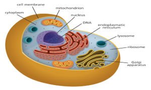
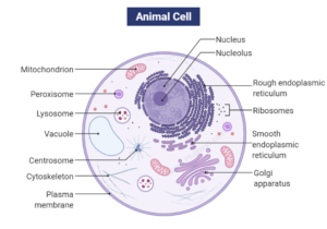
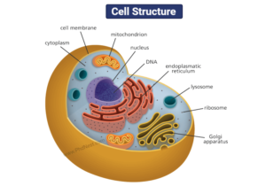
The cell structure comprises individual components with specific functions essential to carry out life’s processes. These components include- cell wall, cell membrane, cytoplasm, nucleus, and cell organelles. Read on to explore more insights on cell structure and function
Cell membrane–
- Lipids + proteins
- Lipids – Phospholipids in the form of bilayer – contain cholesterol.
- Polar head- hydrophilic (loving water) – Outer side; tail – hydrophobic (water hater)– inner side.
- Human beings – In erythrocyte, 52% protein + 40% lipid.
- Peripheral protein – On the surface of membrane; integral – partially or totally buried.
- Fluid mosaic model (1972) – Singer and Nicolson.
Quasi fluid nature of lipid helps in the lateral movement of protein.
- Passive transport – Movement of molecule across P.M. without energy.
- Neutral solute – Simple diffusion (higher concentration- lower)
- Water- osmosis; polar molecule requires carrier protein as they can’t pass through the non-polar lipid bilayer.
- Active transport– ions are transported against the concentration gradient (lower to higher), energy-dependent, ATP utilized. E.g.- Na+/K+ Pump.
Cell wall
- Non – living rigid Structure.
- Algae – composed of cellulose, galactans, mannars, CaCo3.
- Plants – Cellulose, hemicellulose, pectin, protein.
- Primary wall – Capable of growth.
- Secondary wall – Inner side of the cell.
- Middle lamella – Calcium pectate, joins neighboring cell.
- Cell wall & middle lamella is transversed by plasmodesmata.
Endomembrane system
E.g., ER, Golgi complex, lysosome, vacuole.
Endoplasmic reticulum – Network of tiny tubular Structure in the cytoplasm.
- 2 compartment – laminar (inside ER) & extraluminal (cytoplasm).
- RER – Rough Endoplasmic reticulum, bear ribosomes, protein synthesis.
- SER – Smooth, don’t have Ribosomes, lipid synthesis, in animals, they also synthesis steroidal hormones.
Golgi apparatus – Densely stained reticular structures.
- Discovered by Camillo Golgi.
- Contains disc shaped sacs – cisternal (0.5 µm -1 µm) stacked parallel.
- Near nucleus – convex cis/forming face; another side- concave trans/ maturing face.
- Function packaging material, formation of glycoprotein & glycolipids.
- Materials in the form of vesicles from E.R. fuse with cis face and move towards maturing face.
Lysosome
formed by Golgi apparatus, membrane bound.
- Isolated vesicles – rich in a hydrolytic enzyme (lipase, protease, carbohydrate) active at acidic P.H., digest carbohydrate, protein, lipid, a nucleic acid.
Vacuoles – contain water, sap, excretory product not useful by cell.
- Single membrane – tonoplast – facilitate the transport of ions against the concentration gradient, provide buoyancy.
- Amoeba – Contractile vacuole – Excretion; Plants – vacuole (90%).
- Proteins – food vacuole – engulfing food particles.
Mitochondria –
- Sausage shaped of diameter 0.2 – 1 µm.
- Double membranous structure.
- Inner compartment – Homogenous substance – matrix, have infoldings to increase surface area – cristae (for respiration)
- Sites of aerobic respiration produce energy in the form of ATP.
- The powerhouse of the cell, divide by fission.
- Contain own circular DNA, RNA, ribosome (70s)
Plastids –
- Plant cells & Euglenoids.
- On type of pigment – chloroplast, chromoplast and leucoplast.
- Chloroplast – chlorophyll, carotenoid -trap light energy.
- Chromoplast – fat-soluble carotenoid (carotene, xanthophyll) – provides the color.
- Leucoplast – Colorless
- Amyloplast – Store carbohydrates. E.g., Potato.
- Elaioplast – Store oil and fats; Aleuroplast – store proteins.
- Majority of chloroplast – Mesophyll (lens-shaped, discoid)
- One chloroplast per cell – Chlamydomonas; 20-40 per cell – mesophyll.
- Double membrane, inner is less permeable, own circular DNA, 70s ribosome.
- Contain matrix – Stroma which has flattened membranous sacs called thalloid, which are arranged in stacks called grana and connected by stroma lamellae.
- The membrane of the thylakoid contains lumen.
- The stroma contains enzymes for the synthesis of carbohydrates & proteins.
- Light reaction(thylakoid)depends on light
- Dark (stroma) – depends on the product of the light reaction.
Ribosome –
- Granular Structure by George palates.
- Composed of RNA and proteins.
- Has 2 subunit (larger + smaller), S = Svedberg’s unit, sedimentation coefficient.
- Indirectly a measure of density and size.
Cytoskeleton –
- Network of filamentous proteinaceous Structure.
- Provide mechanical support, motility, maintenance of the shape of a cell.
Cilia and Flagella –
- Hair like outgrowth.
- Cilia – Small Structure working like oars, causing movement of the cell.
- Flagella – Comparatively longer and responsible for cell movement.
- Covered with cell membrane, core – axoneme (possess microtubule running parallel to long axis)
- Axoneme has nine pairs of doublets of radially arranged peripheral microtubules and pair of centrally located microtubules – 9 +2.
- The central tubule is connected by bridges enclosed by a central sheath, which is connected to tubules of each doublet (peripheral) by radial spore (9). Peripheral doublets are interconnected by linkers.
- Cilia & flagella emerge from centriole-like Structure – basal bodies.
Centrosome & centrioles –
- Centrosome – Contain 2 centrioles –
- Surrounded by amorphous pericentriolar material.
- Centriole has a cartwheel appearance.
- Made of a peripheral fibril (tubulin protein). Each fibril is a triplet. All are interconnected.
- A central part of centriole is proteinaceous – hub connected triplets by radial spokes made of protein.
- Give rise to spindle apparatus during cell division in animals.
Nucleus –
- Robert Brown (1831)
- Stained by basic dyes by Flemming – chromatin.
- The interphase nucleus (when it is not dividing) has extended and elaborate nucleoprotein fibers – chromatin.
- Nuclear envelop has two parallel membranes with space between them – perinuclear space. The outer membrane remains continuous with the endoplasmic reticulum.
- The membrane is interrupted by small pores, which help in the movement of RNA and protein molecules.
- Erythrocytes and sieve tube cells – enucleated.
- Nucleoplasm – Nucleolus (spherical structure) + chromatin.
- In some stages, cells show structured chromosomes instead of chromatin.
- Chromatin = DNA + protein(histones) +RNA + non histone proteins.
- The human cell has a 2 m long DNA thread among 46 chromosomes.
- Every chromosome has primary constrictions or centromere, which have disc-shaped Structure- kinetochore on sides.
- The centromere holds two chromatids.
On the basis of the position of centromere –
- Metacentric – Middle centromere forming two equal arms of the chromosome.
- Submetacentric – Centromere slightly away from middle resulting in 1 longer and one shorter arm.
- Telocentric – Terminal centromere
Satellite – few chromosomes have non-staining secondary constrictions at const. location giving the appearance of a small fragment.
Microbodies – Membrane-bound minute vesicles that contain enzymes present in both animal and plant cells.
Related Posts
- Phylum Porifera: Classification, Characteristics, Examples
- Dissecting Microscope (Stereo Microscope) Definition, Principle, Uses, Parts
- Epithelial Tissue Vs Connective Tissue: Definition, 16+ Differences, Examples
- 29+ Differences Between Arteries and Veins
- 31+ Differences Between DNA and RNA (DNA vs RNA)
- Eukaryotic Cells: Definition, Parts, Structure, Examples
- Centrifugal Force: Definition, Principle, Formula, Examples
- Asexual Vs Sexual Reproduction: Overview, 18+ Differences, Examples
- Glandular Epithelium: Location, Structure, Functions, Examples
- 25+ Differences between Invertebrates and Vertebrates
- Lineweaver–Burk Plot
- Cilia and Flagella: Definition, Structure, Functions and Diagram
- P-value: Definition, Formula, Table and Calculation
- Nucleosome Model of Chromosome
- Northern Blot: Overview, Principle, Procedure and Results

